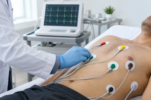

A 2D Echo allows doctors to observe the heart while it is working. Unlike an ECG, which records electrical activity, echocardiography uses ultrasound to create moving images of cardiac structures.
For patients in Kalyan and surrounding areas of Maharashtra, the test may be recommended when a cardiologist needs more information about heart pumping, valves, chambers or other structural features.
What Can a 2D Echo Show?
The heart contains chambers, valves and muscular walls that work together to maintain circulation. A 2D echocardiogram provides real-time images that help a cardiologist evaluate these structures.
Depending on the clinical requirement, the examination can provide information about:
- Heart chamber size
- Heart muscle movement
- Overall pumping function
- Valve structure and movement
- Certain structural abnormalities
- Fluid around the heart
- Selected pressure and blood-flow characteristics when Doppler assessment is included
This makes echocardiography useful across a wide range of cardiac evaluations.
Why Might Your Doctor Recommend It?
A 2D Echo may be considered when symptoms or examination findings suggest that the structure or pumping performance of the heart needs evaluation.
Examples include unexplained breathlessness, swelling in the legs, certain abnormal heart sounds, suspected valve disease or signs that could be associated with reduced cardiac function.
People who already have diagnosed cardiovascular conditions may also undergo repeat echocardiography when their cardiologist considers it clinically necessary.
Is 2D Echo the Same as ECG?
They answer different questions.
An ECG examines the heart's electrical activity. A 2D Echo provides images of the heart's structure and movement.
A person can therefore have an ECG and an echocardiogram during the same cardiac evaluation without the tests being redundant.
For example, someone experiencing palpitations may need an electrical assessment, while breathlessness accompanied by suspected structural disease may require echocardiography. The final investigation plan depends on the complete clinical picture.
What Is Ejection Fraction?
Patients undergoing echocardiography often hear the term ejection fraction or EF.
It is a measurement used to describe how much blood the left ventricle pumps out during contraction. It can provide useful information about left ventricular pumping function, but it should not be interpreted independently.
A cardiologist considers EF alongside symptoms, heart structure, valve findings and other clinical information.
Preparing for a Routine 2D Echo
For a standard transthoracic echocardiogram, extensive preparation is usually unnecessary unless the healthcare team gives specific instructions.
The patient generally lies on an examination couch while a probe is moved over different areas of the chest using ultrasound gel.
The images are obtained from several views so that different cardiac structures can be assessed.
What Happens After the Test?
The important stage is clinical interpretation.
At Muktai Heart Care Clinic, patients undergoing cardiac assessment in Kalyan may have 2D Echo findings reviewed together with symptoms, medical history and other investigations where appropriate.
An abnormal finding does not automatically indicate a severe condition, just as an individual normal measurement does not answer every cardiac question.
Echocardiography is most useful when it addresses a specific clinical concern. By showing how the heart looks and moves, a 2D Echo can help cardiologists determine whether additional monitoring, treatment or investigation should be considered.
Keywords