
Muktai Heart Care Clinic offers comprehensive cardiac evaluation, including 2D Echo, to help diagnose structural and functional heart conditions accurately. While an ECG records the heart's electrical activity, a 2D Echo provides real-time images of the heart, allowing cardiologists to assess how well it is pumping and whether its valves and chambers are functioning normally.
Many people assume that an ECG alone can identify every heart problem. In reality, the two tests serve different purposes and are often recommended together to provide a more complete picture of heart health.
What Is a 2D Echo?
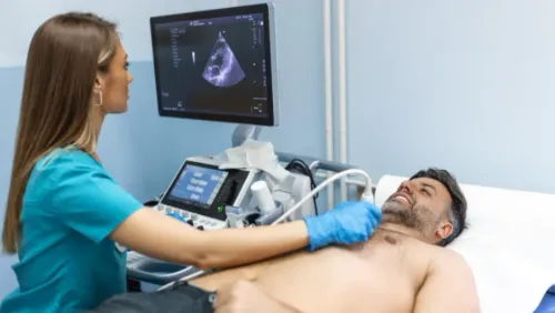
A 2D Echocardiogram, commonly called a 2D Echo, is an ultrasound examination of the heart. It uses sound waves to create moving images without exposing the patient to radiation.
During the procedure, a small handheld device called a transducer is moved over the chest after applying a special gel. The test is painless, non-invasive, and generally completed within 20 to 30 minutes.
What Can a 2D Echo Detect?
A 2D Echo provides detailed information about:
- Heart chamber size
- Pumping efficiency of the heart
- Heart valve function
- Congenital heart abnormalities
- Fluid around the heart
- Thickening of the heart muscle
- Blood flow through the heart using Doppler imaging
This information helps cardiologists understand whether symptoms are caused by structural heart disease rather than electrical abnormalities.
ECG vs 2D Echo: Understanding the Difference
Although both are important cardiac investigations, they answer different clinical questions.
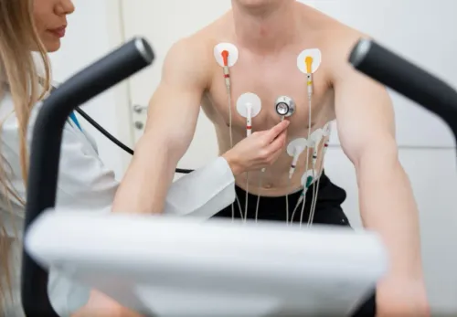
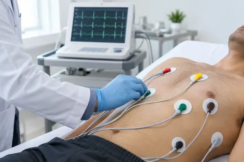
An ECG helps identify:
- Heart rhythm abnormalities
- Heart rate changes
- Evidence of previous or ongoing heart injury
- Electrical conduction problems
A 2D Echo helps evaluate:
- Heart muscle movement
- Valve disorders
- Pumping capacity
- Structural defects
- Blood flow patterns
Depending on the patient's symptoms, one or both tests may be recommended.
When Might Your Doctor Recommend a 2D Echo?
A cardiologist may advise this investigation if you have:
- Chest pain
- Breathlessness
- Swelling in the legs
- Heart murmurs
- High blood pressure affecting the heart
- Suspected valve disease
- Cardiomyopathy
- Follow-up after a heart attack
The test is also commonly used to monitor existing heart conditions over time.
Why Early Imaging Matters
Identifying structural heart problems early allows doctors to:
- Plan appropriate treatment
- Monitor disease progression
- Decide whether medication is sufficient
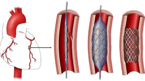
- Evaluate the need for interventional procedures
- Reduce the likelihood of future complications
Because many heart diseases develop gradually, imaging plays a key role in timely diagnosis.
Heart Imaging Services in Kalyan
With increasing awareness about preventive heart care in Kalyan and across Maharashtra, more people are undergoing cardiac evaluation before symptoms become severe. Individuals with diabetes, hypertension, obesity, or a family history of heart disease often benefit from periodic cardiac assessment.
When recommended by a cardiologist, a 2D Echo provides valuable insights into heart function and supports informed decisions about ongoing cardiovascular care.
Keywords